Electrophysiologists partner closely with other cardiologists and cardiac surgeons to achieve the best outcomes for complex cases. Our Electrophysiology Service offers a wide range of diagnostic and treatment options for children — and adults — who suffer from an abnormal heart rhythm.
Popular Search Terms
While the treatment of congenital heart disease (CHD) continues to advance, children are still at risk of developing arrhythmias. An arrhythmia — an abnormal heart rhythm that can make the heart pump inefficiently — can be caused by a congenital heart condition, but it can also be the result of surgery for CHD.
The Surgical Electrophysiology Program at the Benderson Family Heart Center at Boston Children’s was created to find new ways to treat children with CHD and arrhythmias.
We develop strategies to decrease the risk of heart block during CHD surgery. Heart block is an interruption of electrical signals within the heart that causes it to beat irregularly or unpredictably. Surgeons cannot see the conduction tissue that generates the electrical signals because, to the human eye, it looks the same as the rest of the heart muscle. Heart block that occurs during surgery is usually the result of an inadvertent injury.
Innovating to reduce heart block
Our program is a collaboration of the Benderson Family Heart Center’s Department of Cardiac Surgery and the Electrophysiology Service within the Department of Cardiology. Our collective experience and appreciation for innovation is driving us to pinpoint conduction tissue during surgery so that the risk of injuring it and causing heart block in CHD patients is minimized — and will hopefully one day be avoided entirely.
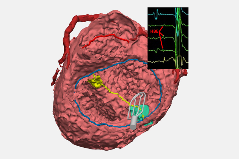
One such recent advancement is already reducing heart block. Our team has developed a protocol for using an electrophysiology (EP) catheter that collects electrical signals from the electrical conduction system of open, beating hearts during surgery. The electrophysiologist in the operating room interprets these signals and communicates the location of conduction tissue back to the surgeons, who then mark the spot of the tissue to avoid inadvertent injury while operating.
Our innovation has reduced heart block in many CHD surgical patients — especially in heterotaxy patients who undergo complex biventricular repair. In that patient group, the incidence of heart block has decreased from more than 14 percent to less than 3 percent when conduction mapping is used during heart surgery.
Gaining an understanding of each child’s heart
When children develop heart block, they often need a pacemaker, committing them to more operations, to maintain the pacemaker’s function, for the rest of their lives. Our ability to map the heart’s conduction system allows us to finally “see” where this tissue is located and give patients a vastly better chance of avoiding surgical complications, so they can focus on recovering from CHD treatment.
EP mapping also allows us to record the location of conduction tissue for further study. We’re finding that anatomical variables — like how a patient’s ventricles are arranged, where the heart is located inside the chest, and how the atria are oriented — can influence the location of the conduction system. While some of these findings were first discovered in older anatomy studies, our rapidly growing conduction mapping experience is enabling us to better understand where the conduction system is located across all variants of complex CHD.
Our program is also creating 3-D models of the cardiac anatomies of every CHD patient who benefits from EP mapping in surgery. Ultimately, those individual models will be available in a digital library that will let clinicians review the influences that different anatomies have on conduction, which may help cardiologists and surgeons developed surgical plans informed by the conduction’s system location for each particular type of CHD.
Mapping conduction tissue is just the first of many innovations and collaborations we intend to pursue in the future, as we collectively work to better treat children with CHD and aim to reduce or eliminate altogether their risk of acquiring arrhythmia.
Electrophysiology surgical mapping volumes
Electrophysiology (EP) mapping has become a routine part of complex congenital heart surgery at Boston Children’s. Pinpointing unseen conduction tissue has led to a significant decrease in heart block following surgery.
Data as of July 2024